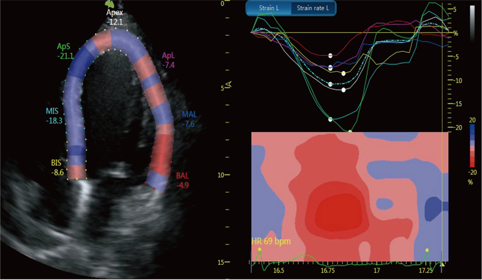
Strain Rate Imaging
Strain Imaging is the quantification of myocardial deformation, either using Tissue Doppler Imaging (TDI) or Speckle Tracking on 2D image (2DTT). Each sample point is tracked frame-to-frame over the cardiac cycle, their dimensional spatio-temporal displacement information is computed to obtain Strain. Positive Strain values are lengthening, thickening, or clockwise rotation. Negative Strain values are shortening, thinning or counter-clockwise rotation. The benefits of strain imaging technology is its ability to differentiate between active and passive movement of myocardial segments, to quantify intraventricular dyssynchrony and to evaluate components of myocardial function, such as longitudinal myocardial shortening, that are not visually assessable, it allows comprehensive assessment of myocardial function and the spectrum of potential clinical applications is very wide.
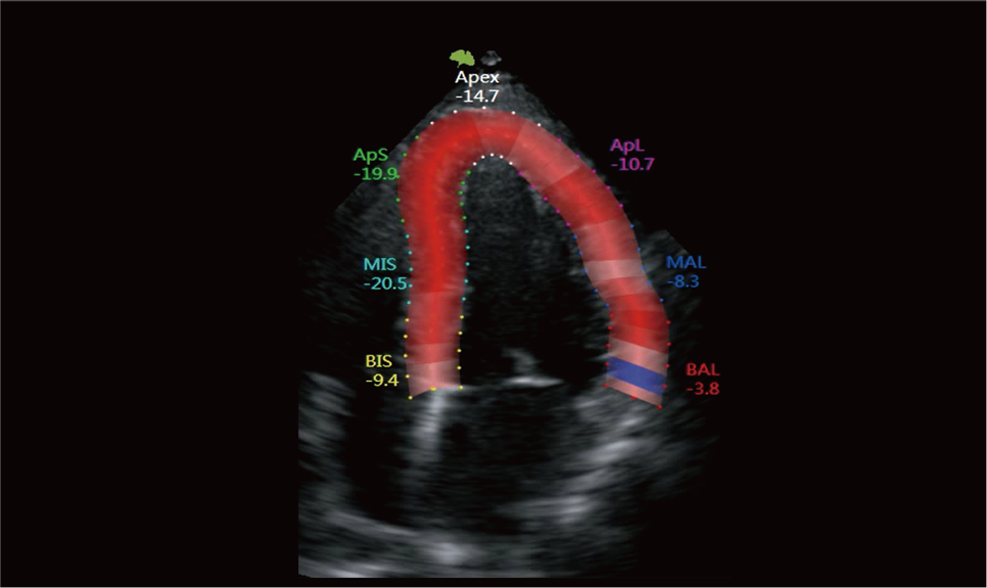
 The preferred ultrasound index to identify movement abnormalities of myocardial segments
Stress Echo
Stress Echocardiography is a non-invasive, dynamic evaluation of myocardial structure and its function under an external stress (exercise or pharmacology). External stresses increase the regional wall motions and thickening thereby increases 3-5 times blood supply to the myocardium to meet the demand in normal individuals. The non-ischaemic dilated or Hypertrophic Cardiomyopathy patients will have less demand due to pathology and ischaemia leads to regional wall motion dysfunction. Thereby, stress echo helps in functional assessment [differentiating active movement from passive/tethering] and structural changes [differentiation thickening/scar/aneurysm- regional bulging] which are more pronounced during stress, can be visually assessed and objectively observed [less subjective compared to standard Echo]. A baseline scan is compared to peak/stages of stress and a standardized wall motion score is performed. Basically, two fundamental types of information can be gathered: tissue viability and coronary flow reserve


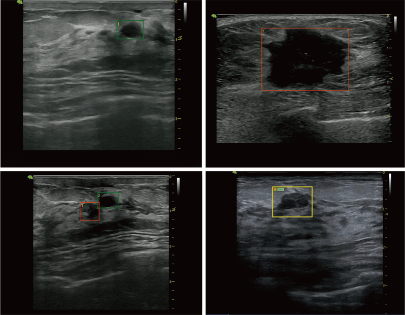 VAid Technology, Real-time Auto Breast Lesion Detection